
Research Interests:
Contents: To investigate the effect of microbial secondary metabolites impinging on cellular signaling, autophagy, apoptosis, programmed necrosis, and to clarify the association among these processes.
Objective: To investigate the new target of the drugs that treated the tumor,and screen out the drugs which could be used in the clinical oncotherapy from microbial secondary metabolites.
The major research achievements:
In the current work, we found that 11'-deoxyverticillin A (C42), a member of the fungal secondary metabolites epipolythiodioxopiperazines (ETPs), could trigger both cell autophagy and apoptosis. However, autophagy precedes apoptotic cell death. C42 could enhance the regulation of receptor-interacting protein 1 (RIP-1) and poly ADP-ribose polymerase (PARP). In another study, it is demonstrated that nuclear factor kappa B (NFkB) could affects autophagy induced by C42.
Representative Pictures:
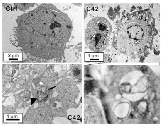
Electron microscopy was performed on vehicle- (Ctrl) and C42-treated (0.25 μM, 6 h) HCT116 cells to detect the autophagosome formation. White arrow heads: the condensation and margination of the chromatin on nuclear membrane; left of low panel: swollen and degenerating mitochondria (white arrows), a vacuole with mitochondria inside (black arrow), a segment of double-membrane structure between a vacuole and mitochondrion (black arrow head); right of low panel: high-contrast image of the cell region indicated by white rectangle.

HCT116 cells were transfected with a plasmid expressing GFP-LC3. After 24 h, the cells were incubated for 6 h at 37°C in DMEM medium with DMSO (Ctrl) or C42 (0.25 μM). Following fixation, cells were stained with DAPI and visualized by confocal microscopy. In contrast to the C42-exposed cells, which showed increased punctate staining of GFP-LC3, GFP-LC3 staining remained diffuse in the control cells.
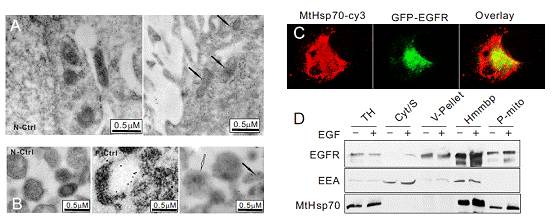
Immune electron microscopy was performed A431 cells (A) or isolated mitochondria of A431 cells (B) to detect the mitochondrial localized EGFR. Colloidal gold particles, which represent the EGFR, were clearly located within the mitochondria, while there is no staining in control cell. N-Ctrl: negative control. (C) GFP-EGFR expressing PAE were permeabilized and immunostained with anti-MtHsp70-Cy3, followed with Cy3-labeled secondary antibodies for imaging. The yellow color representes the overlay between red and green fluorescence.
(D) Fractionation of homogenized A431 cells with or without EGF (100 ng/ml, 1 h) and the resulting fractions were immunoblotted with the antibodies indicated. TH, total homogenate; Cyto/S, cytosol/S100; V-pellet, vesicular pellet; Hmmbp, heavy membrane pellet; P-mito: purified mitochondria.
